



L-type calcium channel
L-type calcium channels (CaV1) convert membrane depolarization into Ca2+ influx in excitable cells, supporting contraction, secretion, gene expression, neuronal excitability, synaptic plasticity, and sensory synaptic transmission[1][2]. Mechanistically, Ca2+ entry through these channels activates calpain, CaMKs, calcineurin, and MAPKs, linking localized Ca2+ signals to fast responses such as muscle contraction and longer-term transcriptional regulation[3]. In skeletal muscle, CaV1.1 directly supports excitation-contraction coupling by acting as the voltage sensor for RyR1, and loss of STAC3 disrupts this coupling and prevents contraction[4]. In disease-relevant models, CaV1.3 loss impairs pacemaking and atrioventricular conduction, while CaV1.2 mutations are associated with inherited cardiac arrhythmia syndromes[5][6]. Compared with CaV2 channels that primarily initiate fast synaptic transmission and CaV3 channels that support repetitive firing, CaV1 channels preferentially regulate contraction, secretion, gene expression, and specialized sensory transmission[1]. For experimental applications, dihydropyridine sensitivity enables isoform-specific studies, and CaV1.1e currents increase with Bay K 8644 and decrease with nifedipine[7][8].
Références:
- [1]. Catterall WA. Voltage-gated calcium channels. Cold Spring Harb Perspect Biol. 2011 Aug 1;3(8):a003947. doi: 10.1101/cshperspect.a003947. PMID: 21746798; PMCID: PMC3140680. et al. Voltage-gated calcium channels. Cold Spring Harb Perspect Biol. 2011 Aug 1;3(8):a003947. [Content Brief]
- [2]. Calin-Jageman I, et al. Ca(v)1 L-type Ca2+ channel signaling complexes in neurons. J Neurochem. 2008 May;105(3):573-83. [Content Brief]
- [3]. Jiang R, et al. Effectiveness of exercise on perinatal depression and anxiety symptoms: A network meta-analysis and dose-response analysis. Int J Gynaecol Obstet. 2026 Jun;173(3):1308-1324. [Content Brief]
- [4]. Rufenach B, et al. Structure and function of STAC proteins: Calcium channel modulators and critical components of muscle excitation-contraction coupling. J Biol Chem. 2021 Jul;297(1):100874. [Content Brief]
- [5]. Mesirca P, et al. Functional role of voltage gated Ca(2+) channels in heart automaticity. Front Physiol. 2015 Feb 2;6:19. [Content Brief]
- [6]. Circulation research thematic synopsis: cardiac arrhythmias. Circ Res. 2013 Jun 7;112(12):e157-79. doi: 10.1161/CIRCRESAHA.113.301756. PMID: 23743230. et al. Circ Res. 2013 Jun 7;112(12):e157-79. [Content Brief]
- [7]. Bourinet E, et al. Dissecting the functional role of different isoforms of the L-type Ca2+ channel. J Clin Invest. 2004 May;113(10):1382-4. [Content Brief]
- [8]. Benedetti B, et al. Physiological and pharmacological modulation of the embryonic skeletal muscle calcium channel splice variant CaV1.1e. Biophys J. 2015 Mar 10;108(5):1072-80. [Content Brief]
All Product Categories
-
L-type calcium channel Inhibitors
(71)
View all products
-
L-type calcium channel Agonists
(2)
View all products
-
L-type calcium channel Antagonists
(23)
View all products
-
L-type calcium channel Activators
(6)
View all products
-
L-type calcium channel Modulator
(1)
View all products
-
L-type calcium channel Controls
(3)
View all products
-
L-type calcium channel Produits associés (109)
Produits associés (109)

-
Minocycline hydrochloride
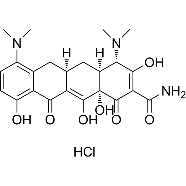 0 ImagesMinocycline hydrochloride is an orally active, potent and BBB-penetrated semi-synthetic tetracycline antibiotic. Minocycline hydrochloride is a hypoxia-inducible factor (HIF)-1α inhibitor. Minocycline hydrochloride shows anti-cancer, anti-inflammatory, and glutamate antagonist effects. Minocycline hydrochloride reduces glutamate neurotransmission and shows neuroprotective properties and antidepressant effects. Minocycline hydrochloride inhibits bacterial protein synthesis through binding with the 30S subunit of the bacterial ribosome, resulting in a bacteriostatic effect.
-
Bay K 8644
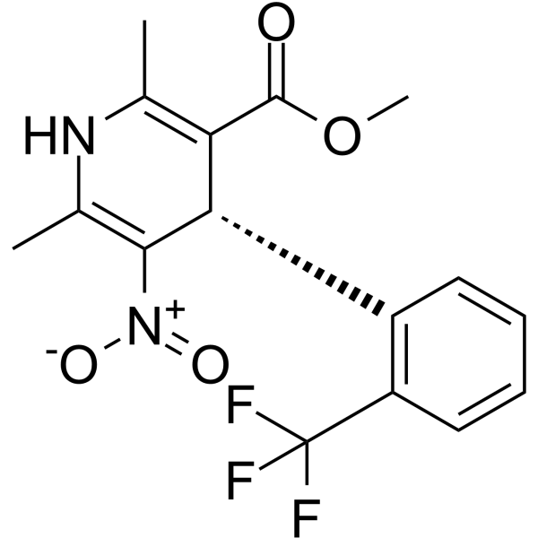
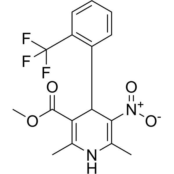 0 ImagesSynonyms: (±)-Bay K 8644Bay K 8644 ((±)-Bay K 8644) is a racemate consisting of two isomers (R)-(+)-Bay-K-8644 and (S)-(-)-Bay-K-8644. Bay K 8644 is a L-type Ca2+ channel agonist with an EC50 of 17.3 nM. Bay K 8644 increases Ca2+ influx through sarcolemmal Ca2+ channels by increasing the open time of the channel. Bay K 8644 has vasoconstrictive effects.
-
Ruthenium red
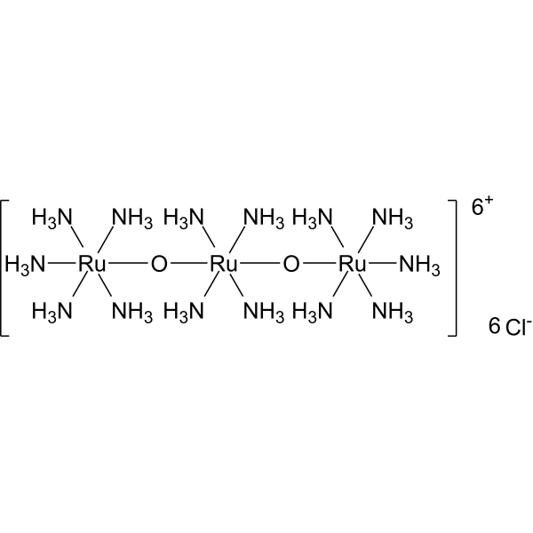 0 ImagesSynonyms: Ammoniated ruthenium oxychlorideRuthenium red (Ammoniated ruthenium oxychloride) is a polycationic dye widely used for electron microscopy (EM) of cells, tissues and vegetative bacteria. Ruthenium red strongly reacts with phospholipids and fatty acids and binds to acidic mucopolysaccharides. Ruthenium red is a L-type calcium current (ICa) blocker. When using Ruthenium Red to stain plant tissue sections affected by pathogens, the breakdown of mucilaginous substances by the pathogens prevents them from taking up the stain, whereas the cell walls of healthy tissue stain red.
-
Amlodipine
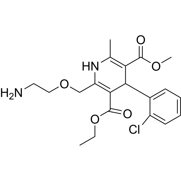 0 ImagesAmlodipine, an antianginal agent and an orally active dihydropyridine calcium channel blocker, works by blocking the voltage-dependent L-type calcium channels, thereby inhibiting the initial influx of calcium. Amlodipine can be used for the research of high blood pressure and cancer.
-
Ethacrynic acid
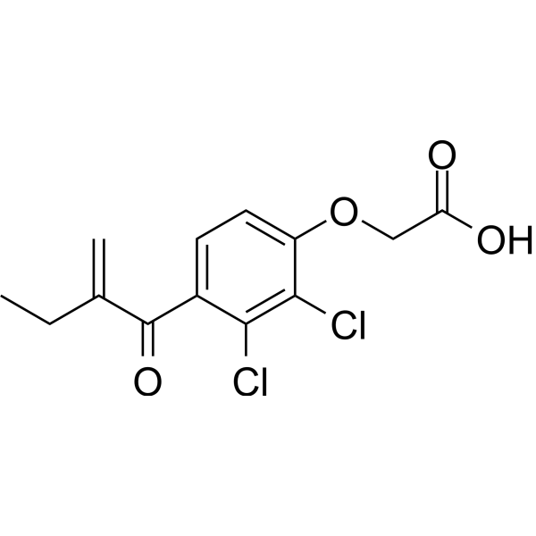 0 ImagesEthacrynic acid has anti-inflammatory and anticancer activity. Ethacrynic acid is an orally active diuretic. Ethacrynic acid is an inhibitor of glutathione S-transferase (GSTs) and Wnt signaling pathways. Ethacrynic acid is a radiosensitizer. Ethacrynic acid can inhibit airway smooth muscle (ASM) contraction in mice. Ethacrynic acid can increase the outflow of aqueous humor from the eye for the study of glaucoma.
-
Bepridil hydrochloride
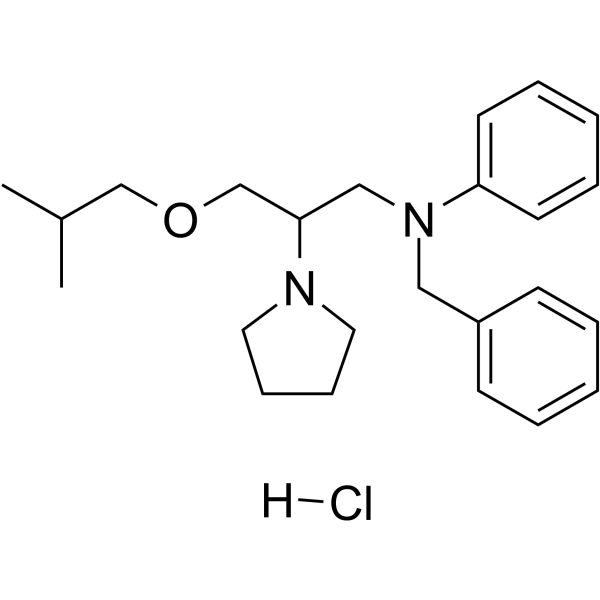 0 ImagesSynonyms: CERM 1978; Org 5730 hydrochlorideBepridil hydrochloride (CERM 1978; Org 5730 hydrochloride) is an orally active non-selective calcium channel antagonist with multi-ion channel blocking activity. Bepridil hydrochloride modulates Calmodulin, the 20S proteasome, T-type/L-type calcium channels, cardiac sodium channels, multiple potassium channels, γ-secretase, β-secretase, and mitoKATP/sarcKATP channels. Bepridil hydrochloride acts as a hydroxyl radical scavenger, regulates mitochondrial and intracellular calcium handling, and exerts antiarrhythmic, antianginal, and cardioprotective effects. Bepridil hydrochloride alters amyloid precursor protein processing, reduces β-amyloid and thalamic calcium levels, restores seladin-1/DHCR24 expression, and improves sensorimotor recovery after cerebral ischemia. Bepridil hydrochloride also exhibits potent inhibitory effects on SARS-CoV-2 replication. Bepridil hydrochloride is used in studies related to stable angina, arrhythmias, cerebral ischemia, Alzheimer's disease, and SARS-CoV-2 infection.
-
Nisoldipine
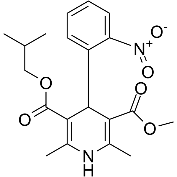 0 ImagesSynonyms: BAY-k 5552Nisoldipine (BAY-k 5552; Sular) is an orally active and blood-brain barrier-penetrating dihydropyridine calcium antagonist, with greater vascular selectivity than other calcium channel antagonists. Nisoldipine inhibits calcium influx and blocks voltage-gated calcium channels. Nisoldipine dilates coronary and systemic arteries. Nisoldipine has antihypertensive and anti-anginal activity. Nisoldipine also displays neuroprotective and antiviral activity.
-
Diltiazem
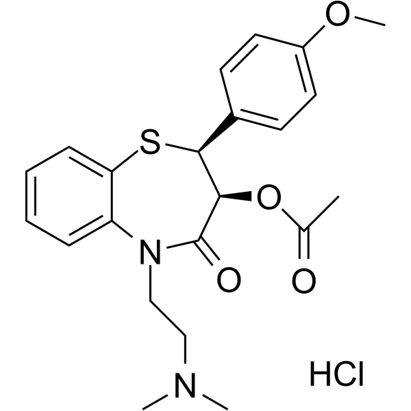
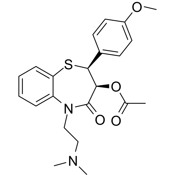 0 ImagesDiltiazem is an orally active L-type Ca2+ channel blocker. Diltiazem shows antihypertensive and antiarrhythmic effects. Diltiazem can be used for the research of cardiac arrhythmia, hypertension, and angina pectoris.
-
Flufenamic acid
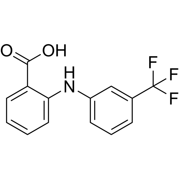 0 ImagesFlufenamic acid is a non-steroidal anti-inflammatory agent, inhibits cyclooxygenase (COX), activates AMPK, and also modulates ion channels, blocking chloride channels and L-type Ca2+ channels, modulating non-selective cation channels (NSC), activating K+ channels. Flufenamic acid binds to the central pocket of TEAD2 YBD and inhibits both TEAD function and TEAD-YAP-dependent processes, such as cell migration and proliferation.
-
Isradipine
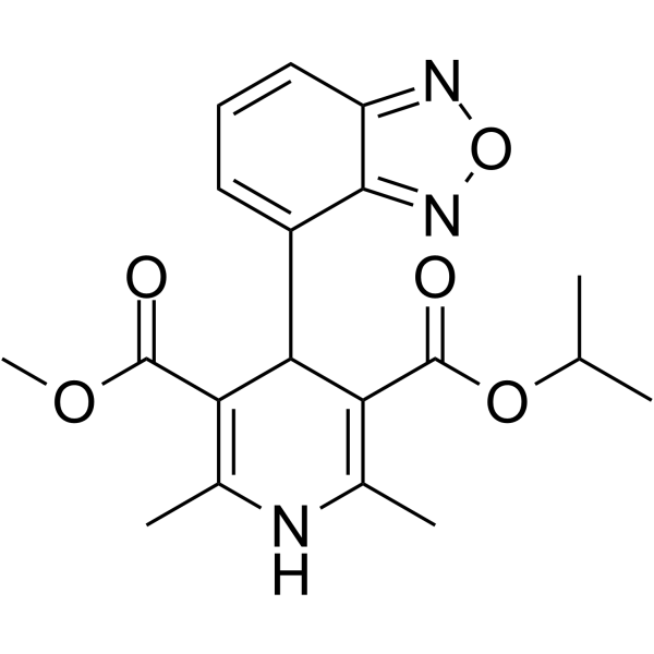 0 ImagesSynonyms: PN 200-110Isradipine (PN 200-110) is an orally active and blood-brain barrier permeability L-type calcium channel blocker. Isradipine, as a powerful peripheral vasodilator, is a dihydropyridine calcium antagonist with selective actions on the heart as well as the peripheral circulation. Isradipine is a potentially viable neuroprotective agent for Parkinson's disease.
-
Cilnidipine
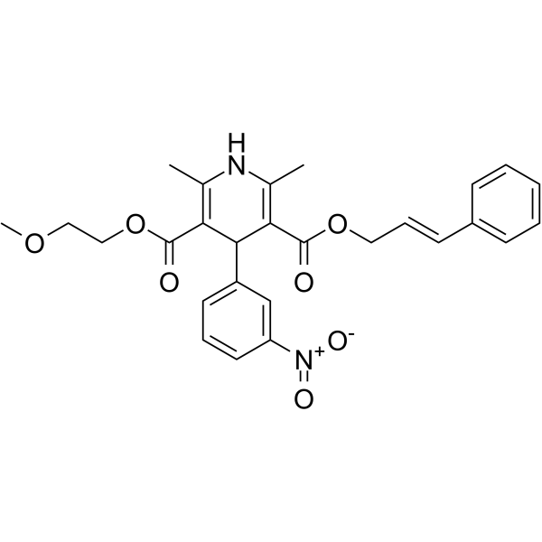 0 ImagesSynonyms: FRC-8653
-
Felodipine
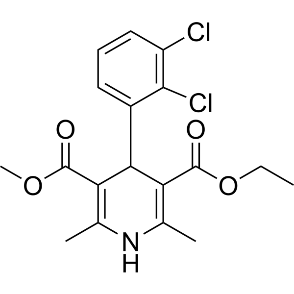 0 ImagesFelodipine, a dihydropyridine, is a potent, vasoselective calcium channel antagonist. Felodipine lowers blood pressure (BP) by selective action on vascular smooth muscle, especially in the resistance vessels. Felodipine, an anti-hypertensive agent, induces autophagy. Felodipine can cross the blood-brain barrier.
-
Urolithin C
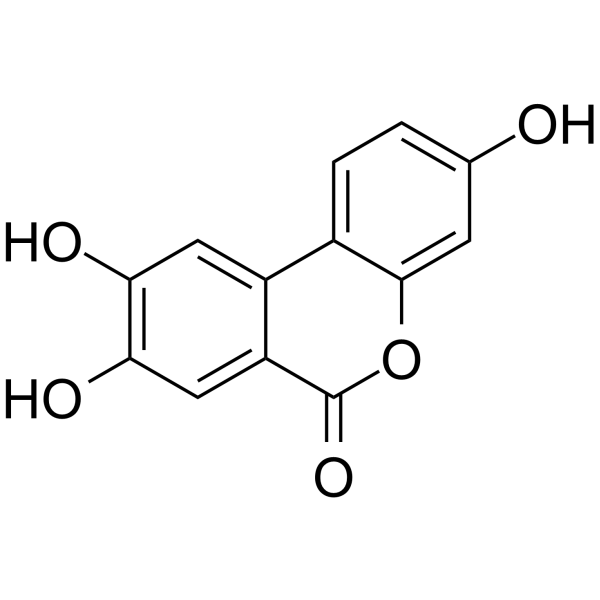 0 ImagesUrolithin C, a gut-microbial metabolite of Ellagic acid, is a glucose-dependent activator of insulin secretion. Urolithin C is a L-type Ca2+ channel opener and enhances Ca2+ influx. Urolithin C induces cell apoptosis through a mitochondria-mediated pathway and also stimulates reactive oxygen species (ROS) formation.
-
Ginsenoside Rd
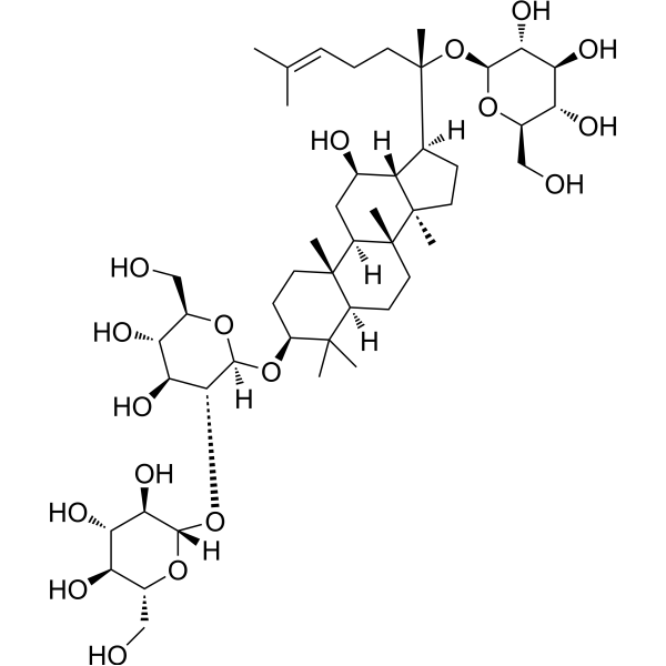 0 ImagesSynonyms: Gypenoside VIIIGinsenoside Rd inhibits TNFα-induced NF-κB transcriptional activity with an IC50 of 12.05±0.82 μM in HepG2 cells. Ginsenoside Rd inhibits expression of COX-2 and iNOS mRNA. Ginsenoside Rd also inhibits Ca2+ influx. Ginsenoside Rd inhibits CYP2D6, CYP1A2, CYP3A4, and CYP2C9, with IC50s of 58.0±4.5 μM, 78.4±5.3 μM, 81.7±2.6 μM, and 85.1±9.1 μM, respectively.
-
Amlodipine besylate
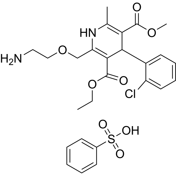 0 ImagesSynonyms: Amlodipine benzenesulfonateAmlodipine besylate (Amlodipine benzenesulfonate), an antianginal agent and an orally active dihydropyridine calcium channel blocker, works by blocking the voltage-dependent L-type calcium channels, thereby inhibiting the initial influx of calcium. Amlodipine besylate can be used for the research of high blood pressure and cancer.
-
Minocycline
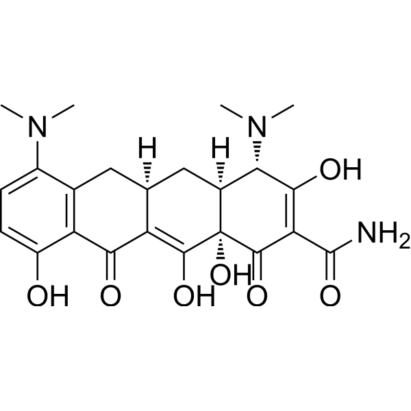 0 ImagesMinocycline is an orally active, potent and BBB-penetrated semi-synthetic tetracycline antibiotic. Minocycline is a hypoxia-inducible factor (HIF)-1α inhibitor. Minocycline shows anti-cancer, anti-inflammatory, and glutamate antagonist effects. Minocycline reduces glutamate neurotransmission and shows neuroprotective properties and antidepressant effects. Minocycline inhibits bacterial protein synthesis through binding with the 30S subunit of the bacterial ribosome, resulting in a bacteriostatic effect.
-
Mibefradil dihydrochloride
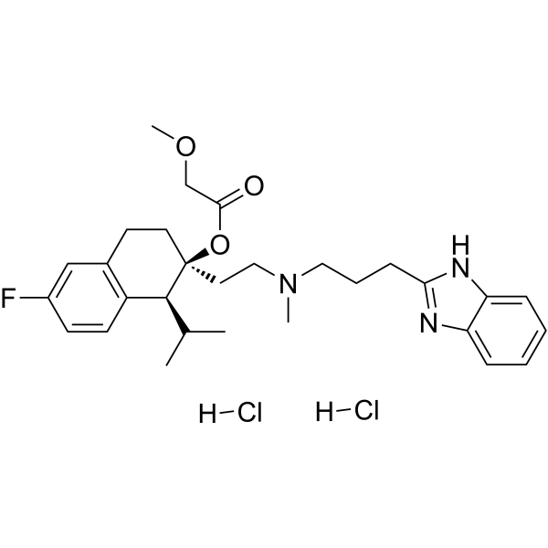 0 ImagesSynonyms: Ro 40-5967 dihydrochlorideMibefradil dihydrochloride (Ro 40-5967 dihydrochloride) is a calcium channel blocker with moderate selectivity for T-type Ca2+ channels (IC50s of 2.7 μM and 18.6 μM for T-type and L-type currents, respectively).
-
Diacerein
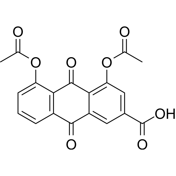 0 ImagesSynonyms: Diacerhein; DiacetylrheinDiacerein (Diacerhein), an orally active anthraquinone, reduces production of IL-1 converting enzyme then inhibits the activation of IL-1β by related downstream signaling. Diacerein is an anti-inflammatory and anti-rheumatic drug. Diacerein can relieve bronchospasm and control airway inflammation in asthmatic mice. Diacerein has the potential for slow acting drug in osteoarthritis (SYSADOA) research.



